Rib Cage Muscles Diagram / Intercostals Rehab My Patient / Rib cage diagram this summary post is displaying rib cage diagram … please click on the diagram(s) to view larger version.
Rib Cage Muscles Diagram / Intercostals Rehab My Patient / Rib cage diagram this summary post is displaying rib cage diagram … please click on the diagram(s) to view larger version.. The last diagram shows how the ribs are connected to the vertebral column or spine. As you inhale, the muscles in between the ribs lift the rib cage up, allowing the lungs to expand. Quickly memorize the terms, phrases and much more. Best viewed on 1280 x 768 px resolution in any modern browser. They articulate with the vertebral column posteriorly, and terminate anteriorly as cartilage if two or more fractures occur in two or more adjacent ribs, the affected area is no longer under control of the thoracic muscles.
These rib muscles automatically get worked when you do bench presses, push ups and dips, but a few bonus exercises can help you really zero in for a more chiseled torso. You'll need a bench and one dumbbell to do this exercise. Start studying rib cage muscles. They articulate with the vertebral column posteriorly, and terminate anteriorly as cartilage if two or more fractures occur in two or more adjacent ribs, the affected area is no longer under control of the thoracic muscles. Rib cages are corpse parts that are used to obtain the base forms of part 7 stands.
Rib Cage Muscles Diagram Rib Muscles Diagram Blank Doing Wiring Diagram The New Way Posted On December 22 2018december 22 2018 Cendoo from i2.wp.com Anatomical illustrations and diagrams of the spine (cervical, dorsal and lumbar) and back: These muscles may be located anteriorly, posteriorly, and/or laterally. Best viewed on 1280 x 768 px resolution in any modern browser. Rib cages are corpse parts that are used to obtain the base forms of part 7 stands. This item can be dropped. It is formed by the vertebral column, ribs, and sternum and encloses the heart and lungs. The last diagram shows how the ribs are connected to the vertebral column or spine. As a consequence, rib cage expansion predominates during quiet breathing in the seated position and abdominal expansion predominates in the supine position.
The two muscles which comprise the intermediate muscle group are the serratus posterior inferior, and the serratus posterior superior.
During normal breathing, the major inspiratory muscles produce rib cage expansion and a downward movement of the diaphragm. The other attachment of these muscles is usually considered to be either superior or inferior to the rib attachment. You'll need a bench and one dumbbell to do this exercise. Best viewed on 1280 x 768 px resolution in any modern browser. This is an online quiz called rib cage muscle diagram. As you inhale, the muscles in between the ribs lift the rib cage up, allowing the lungs to expand. In humans, the rib cage, also known as the thoracic cage. These muscles may be located anteriorly, posteriorly, and/or laterally. Study flashcards on chapter 10 muscle diagrams at cram.com. The primary responsibilities of the ribcage involve protecting the thoracic visceral organs, enclosing the thoracic visceral organs, and is included in the general mechanics of the process of breathing. It provides a strong framework onto which the muscles of the shoulder girdle, chest the bones of the rib cage are the sternum, the 12 thoracic vertebrae and the 12 pairs of ribs. At the chest many rib bones connect to the sternum via costal cartilage segments of hyaline cartilage that allow the rib cage to expand during respiration. The muscles you're referring to are called the intercostals, and i've found that they're best built with calisthenics because they're stabilizer muscles.
During normal breathing, the major inspiratory muscles produce rib cage expansion and a downward movement of the diaphragm. The muscles you're referring to are called the intercostals, and i've found that they're best built with calisthenics because they're stabilizer muscles. Anatomical illustrations and diagrams of the spine (cervical, dorsal and lumbar) and back: Learn vocabulary, terms and more with flashcards, games and other study tools. The function of the rib cage is to filter the blood it receives, processing the blood.
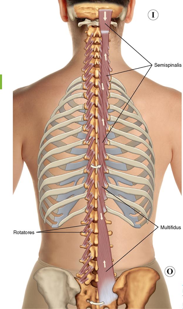
8 Muscles Of The Spine And Rib Cage Musculoskeletal Key from musculoskeletalkey.com The last diagram shows how the ribs are connected to the vertebral column or spine. Start studying rib cage muscles. The rib cage has three important functions: Perform dumbbell pullovers to work the muscles along your rib cage. Learn vocabulary, terms and more with flashcards, games and other study tools. Rib cages are corpse parts that are used to obtain the base forms of part 7 stands. For more anatomy content please follow us and visit our website: Rib cage diagram this summary post is displaying rib cage diagram … please click on the diagram(s) to view larger version.
The function of the rib cage is to filter the blood it receives, processing the blood.
Rib cage diagram this summary post is displaying rib cage diagram … please click on the diagram(s) to view larger version. This is an online quiz called rib cage muscle diagram. Rib cage diagram with organs. Muscles that move the rib cage attach to the rib cage. All muscles that are attached to the human rib cage have the inherent potential to cause a breathing action. Osteology, myology of the spine. These muscles may be located anteriorly, posteriorly, and/or laterally. The blood supply to the tibialis anterior muscle comes primarily from the anterior tibial artery and its branches. Muscles that helpful in expanding the thoracic cavity are called the inspiratory muscles because they help in inhalation, while those that compress the thoracic cavity are called expiratory. Anatomical illustrations and diagrams of the spine (cervical, dorsal and lumbar) and back: You'll need a bench and one dumbbell to do this exercise. Perform dumbbell pullovers to work the muscles along your rib cage. Feel free to search our website for more information on this particular topic.
This is an online quiz called rib cage muscle diagram. Anatomical illustrations and diagrams of the spine (cervical, dorsal and lumbar) and back: Identify muscles of the rib cage. These rib muscles automatically get worked when you do bench presses, push ups and dips, but a few bonus exercises can help you really zero in for a more chiseled torso. At the chest many rib bones connect to the sternum via costal cartilage segments of hyaline cartilage that allow the rib cage to expand during respiration.
1 Intercostal Muscles Diagram Quizlet from o.quizlet.com As a consequence, rib cage expansion predominates during quiet breathing in the seated position and abdominal expansion predominates in the supine position. Start studying rib cage muscles. Learn vocabulary, terms and more with flashcards, games and other study tools. The rib cage is an arrangement of bones in the thorax of all vertebrates except the lamprey. Rib cage diagram this summary post is displaying rib cage diagram … please click on the diagram(s) to view larger version. In humans, the rib cage, also known as the thoracic cage. All muscles that are attached to the human rib cage have the inherent potential to cause a breathing action. Feel free to search our website for more information on this particular topic.
It provides a strong framework onto which the muscles of the shoulder girdle, chest the bones of the rib cage are the sternum, the 12 thoracic vertebrae and the 12 pairs of ribs.
They are somewhat rare, but not too valuable. According to their attachment to the sternum the ribs are classified into three groups. Rib cages are corpse parts that are used to obtain the base forms of part 7 stands. In humans, the rib cage, also known as the thoracic cage. Rib cage diagram this summary post is displaying rib cage diagram … please click on the diagram(s) to view larger version. Study flashcards on chapter 10 muscle diagrams at cram.com. It encloses and protects the heart and lungs. This is an online quiz called rib cage muscle diagram. Muscles that move the rib cage attach to the rib cage. When you exhale, the rib cage moves down again, squeezing the air. The ribs are a set of twelve paired bones which form the protective 'cage' of the thorax. For more anatomy content please follow us and visit our website: Ribs, respiratory muscles and respiratory system | researchgate, the professional network for scientists.


0 Komentar